How the dog neck is anatomically structured
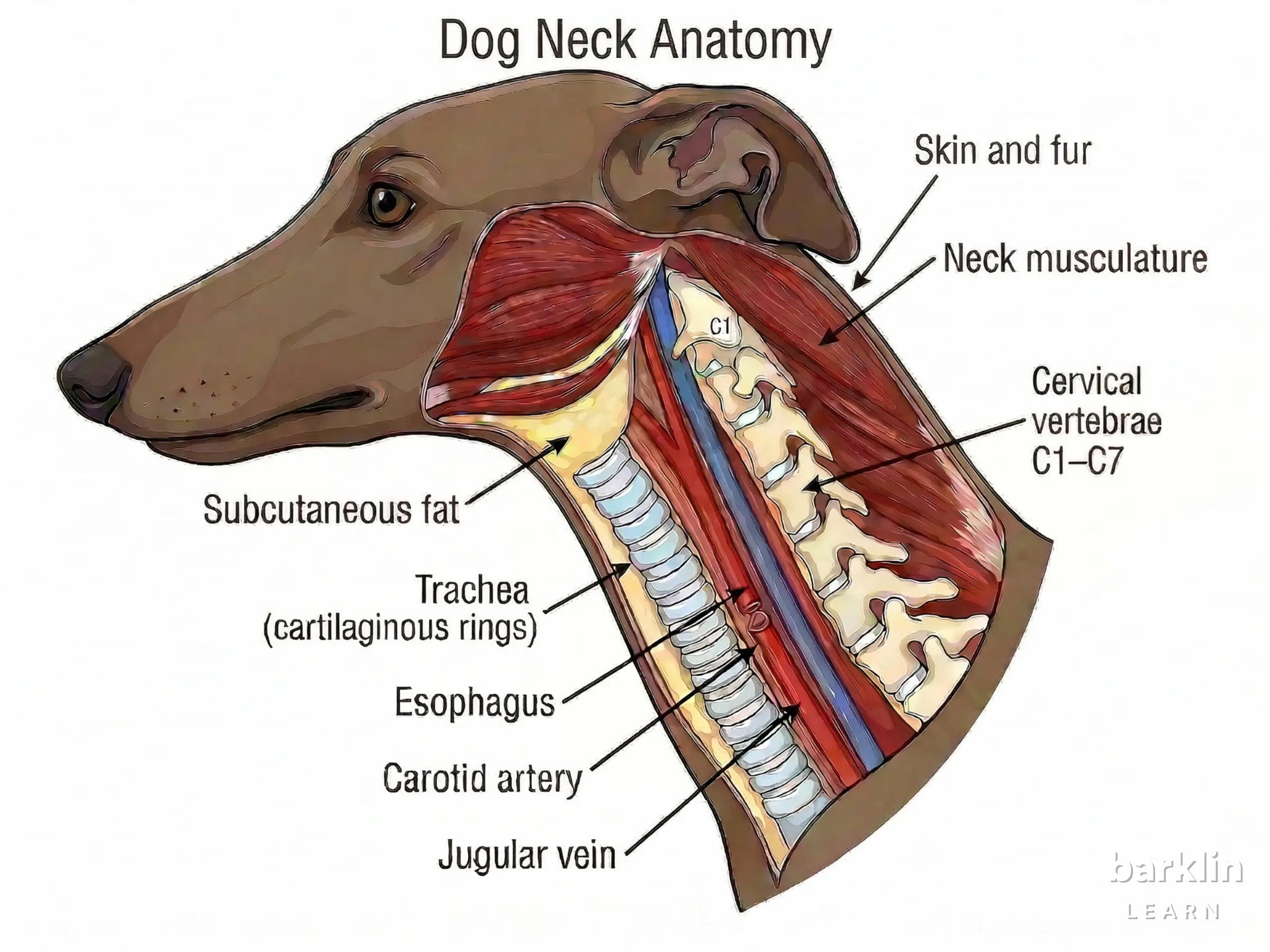
The dog neck is not a uniform cylinder but a layered organ system made up of several mechanically distinct zones. From outside to inside: coat and skin, subcutis as a loose connective-tissue cushion, a superficial fascial layer, neck musculature at two functionally separated depths, trachea and oesophagus as ventral conduction structures, and the carotid sheath (Vagina carotica) with its neurovascular structures, and finally the cervical spine as the deep bony axis.
Seven cervical vertebrae (C1–C7) form this axis. This number applies to every dog, regardless of breed, body size, or neck shape. The vertebrae sit dorsally offset, occupying the neck’s deep posterior side rather than its cross-sectional centre. The trachea and oesophagus run anterior to the bony axis; deeper-lateral sits the carotid sheath. The jugular groove (Sulcus jugularis) with the external jugular vein marks the superficial-lateral zone: the first structure that lateral contact reaches.
Soft, compressible tissue lies at the top; stiff, dense structures lie deeper. This order cannot be bypassed; it is anatomically fixed. And it determines which layer responds mechanically first when surface contact is introduced.
This anatomy forms the reference frame for all later questions about pressure distribution, neck circumference, and the contact geometry of collars. Anyone who needs the wide dog collar as a starting concept will find the structural foundation at Wide dog collar: foundational concept. This page evaluates no products and makes no recommendations; it describes the anatomical basis that downstream pages build on.
Which structures form the cervical axis
The bony cervical axis of the dog consists of seven cervical vertebrae (C1–C7), which divide into two functionally distinct groups. Atlas (C1) is ring-shaped and forms the cranial bony boundary of the cervical canal; it carries the skull and allows nodding. Axis (C2) carries the dens axis as its rotation axis and enables lateral head rotation. C3 through C7 follow as morphologically similar segmental elements and together form the load-bearing cervical axis.
Between the vertebral bodies lie the intervertebral discs, whose nucleus pulposus has a water content of approximately 70–90% and distributes axial load hydromechanically between segments. Laterally through the transverse foramina (C1–C6) runs the vertebral artery. The trachea runs with approximately 35–45 horseshoe-shaped cartilaginous rings as the ventral conduction structure anterior to the axis, in a deep airway space overlaid by multiple tissue and muscle layers, well below the skin surface.
As diagram 2 shows, Atlas (C1) and Axis (C2) are morphologically distinct from the uniform segment sequence of C3–C7.

This skeletal continuity provides the deep reference frame to which all softer cervical structures are topographically related; the bony axis itself does not receive surface load directly, but only after transmission through the overlying layers.
The following table maps the principal cervical structures of the dog neck along the deep axis.
| Structure | Anatomical finding | Mechanical relevance |
|---|---|---|
| Atlas (C1) | ring-shaped geometry | cranial bony boundary |
| Axis (C2) | dens axis as rotation point | centre of movement |
| C3–C7 | segmental sequence | load-bearing cervical axis |
| Intervertebral discs | nucleus pulposus approx. 70–90% water | pressure distribution between segments |
| Trachea | approx. 35–45 cartilaginous rings | ventral conduction structure anterior to the axis |
This makes clear which components define the deep cervical axis and which merely lie anterior to or around it.
How tissue layers transfer force into depth
The depth order of cervical tissue follows a mechanical logic: every externally introduced force reaches the softest, most superficial structures first. Skin and subcutis have a tissue stiffness of approximately 1–10 kPa; the superficial fascia falls in the range of approximately 50–200 kPa. Tracheal cartilage, by contrast, sits in the megapascal range, several orders of magnitude stiffer than all the soft tissue above it.
That is the structural core rule of this model.
If contact is introduced at the neck surface → skin, subcutis, and fascia deform first; deeper airway and skeletal spaces are mechanically engaged only through this layered step-down transfer. If the contact area remains small → local contact pressure in the superficial zone rises; deeper layers only receive increasing load once the deformation capacity there is exhausted. If the contact area is larger → the introduced force distributes across more tissue before structurally rigid compartments such as tracheal cartilage or vertebral bone are mechanically reached.
Contact geometry determines the transfer path.
One model boundary: the anatomical layering does not by itself produce uniform force distribution; that depends on contact geometry, surface area, and local topography.
Neck musculature integrates into this layer system at two functionally separated depths. Diagram 4 shows the neck musculature of the dog in its layers and makes visible how superficial and deep muscle groups take on topographically distinct roles.

Muscle mass creates a topographically graduated buffer: a contoured transition zone with varying depth positions depending on contact site. Superficial muscles such as M. brachiocephalicus and M. sternocephalicus shape the neck’s contour and movement. Deeper muscles (M. splenius, M. semispinalis capitis, M. longus capitis) stabilise the vertebral joints segmentally. Surface contact never reaches a homogeneous muscle wall.
Depth zones of the dog neck
| Zone | Dominant structures | Mechanical role | Helps read |
|---|---|---|---|
| Superficial | Skin, subcutis, jugular groove | first contact and deformation zone | Section 1 and Section 5 |
| Intermediate | Neck musculature and fascia | topographic buffer and force redistribution | Section 3 |
| Deep | Trachea, carotid sheath, cervical spine | airway, vascular, and skeletal axis space | Section 2 and Section 4 |
Which structures lie superficially and deeply in cross section
The cross section at C3 level is the most informative plane for reading the topographic order of the dog neck directly. At this level, the jugular groove and external jugular vein lie in the superficial-lateral zone, the first contact zone for laterally applied force. Below them sits the intermediate zone of neck musculature and fascia, which acts as a topographic buffer between the surface and the deep structures.
Ventrally, the trachea sits as a deep conduction structure; from C3 onwards the oesophagus begins to shift dorsally, displacing toward the left from approximately C4. The carotid sheath, which contains the common carotid artery, internal jugular vein, and vagosympathetic trunk, lies deeper-lateral and is separated from the jugular groove by fascia and muscle tissue. The cervical spine forms the dorsal reference axis. These structures do not all share a single depth plane; cross-sectional anatomy separates them by function and position.
The comparison table shows which structures lie superficially, intermediately, or deeply within the cross section.
| Structural zone | Position at C3 level | Mechanical consequence |
|---|---|---|
| Jugular groove / external jugular vein | superficial-lateral | early-exposed contact zone |
| Neck musculature / fascia | intermediate | force redistribution and topographic buffer |
| Trachea / oesophagus | ventral / dorsolateral offset | deep conduction space, not at surface level |
| Carotid sheath | deep-lateral | neurovascular compartment |
| Cervical spine | dorsal-deep | osseous reference axis |
This makes clear that the same external force meets very different topographic conditions depending on which zone it contacts. The anatomical consequence depends on the position of the contact, not on a universal “neck layer.” Breed differences shift the proportions of these zones, not whether they exist. How these zone proportions shift morphologically in sighthounds is described in Sighthound neck geometry compared.
What this anatomy means for neck contact and contact zones
A collar does not meet a homogeneous cylinder. It meets an anatomically graduated, slightly conical surface whose local contact situation depends on contact width, band position, and the relationship between the superficial tissue layer and deeper-lying structures. The physics of contact follows the basic relation P = F / A: at constant traction force, local contact pressure rises as contact area decreases.
A mean traction force of approximately 30 N serves as the reference value. Local peak pressures can exceed 800 kPa when the contact area is small. A distributed reference value of approximately 0.43 N/cm² provides a biomechanical comparison point for wide-area load distribution. These values describe orders of magnitude. They evaluate no systems and make no recommendations.
As diagram 3 shows at the C3 cross section, the trachea, carotid sheath, and jugular groove lie in clearly separated topographic planes. They do not share a surface.

Contact zone geometry is a depth problem. Which structure becomes mechanically relevant at a given contact width depends on its topographic position. There is no single, generic neck surface. Anatomy explains this relationship; it neither organises product systems nor evaluates constructions.
How a dog collar should fit in practice is described in How a dog collar should fit.
For a deeper understanding of how pressure distribution and collar width relate on the basis of this anatomy, see Understand pressure distribution at the dog neck.
System boundaries
This model describes the static depth order of cervical structures and their topographic relationships. It does not model dynamic load sequences, clinical assessments, or product comparisons.
| Topic out of scope | Further reading |
|---|---|
| Evaluating collar versus harness | Comparing force paths of collar and harness ↗ |